 Topografi
 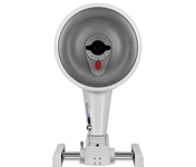 Tomographie
 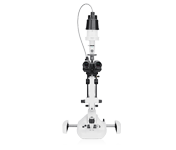 Biyomikroskop Dokümantasyonu
 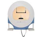 Perimetri
  Tonometre
  Görme Testi Ekipmanı
 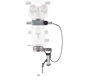 Vitreoretinal Surgery
  Manüel Kırılım
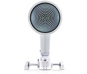
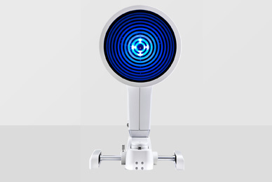
OCULUS Keratograph 5M, yerleşik gerçek keratometreye ve harici görüntüleme için optimize edilmiş renkli bir kameraya sahip bir gelişmiş korneal topograftır. Özgün özelliklere meibomian bezlerinin incelenmesi, non-invazif gözyaşı filmi kırılma süresi ve gözyaşı menisküs yükseklik ölçümü ve lipid katmanı değerlendirmesi dâhildir.

|
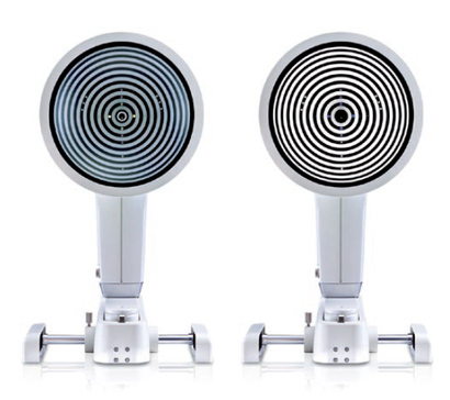
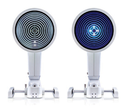
OCULUS Keratograph 5M |


Keratograph 5M ile hastalarınıza daha önce hiç görmedikleri görüntüleri gösterebilirsiniz. Muayeneler ve takipler sırasında profesyonel danışmanlık sağlayarak hastanın güvenini kazanın.

Keratograph 5M'yi konsültasyonunuza aktif bir şekilde entegre edin. Çok sayıdaki kolay anlaşılır ekran, iletişim ve hasta eğitimi konusunda sizi destekler. Hizmetlerinizi şeffaf hale getirmek için Keratograph 5M'nizi bir pazarlama aracı olarak kullanın!
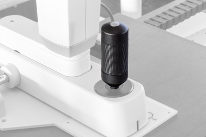
Keratograph 5M için yeni kumanda kolu, doğrudan cihazdan görüntüleri ve video sekanslarını yakalamanızı sağlar. Fare kullanmaya gerek kalmaz: Görüntü veya video yakalarken elleriniz sadece kumanda kolunun üzerinde durur.
Yeni kumanda kolu, akıllı bir Bluetooth arayüzü ile iletişim kurar. Ayrıca, kullanışsız güç kabloları ile uğraşma ihtiyacını ortadan kaldırır. Kumanda kolu kullanılmadığında, pil ömrünü artırmak için uyku modu özelliği devreye girer.
Yeni kumanda koluna geçiş yapmak hızlı ve basittir. Ne kadar kolay olduğunu görmek için aşağıdaki videoyu izleyin!
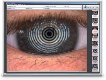
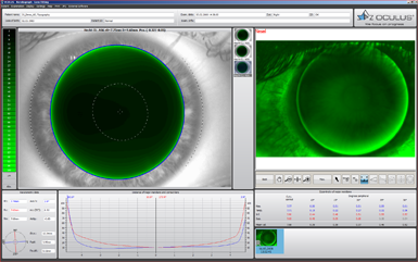
Korneanın tüm yüzeyini ölçmek için binlerce ölçüm noktası kullanılır. Bu amaç için beyaz halka aydınlatma kullanılır. Yansıma etkisine tepki olarak salgılanan gözyaşından bağımsız olarak gözyaşı filminin analizini yapabilmek için bir kızıl ötesi aydınlatma halkası sunulmuştur.
Keratograph 5M'nin her işlevine mükemmel aydınlatma entegre edilmiştir: Gözyaşı filmi dinamikleri için beyaz diyotlar, fluo-görüntüler için mavi diyotlar, Meibografi için kızıl ötesi diyotlar.
 |
| Hassasiyet | ± 0,1 D |
| Tekrarlanabilirlik | ± 0,1 D |
| Halka sayısı | 22 |
| Çalışma mesafesi | 78 - 100 mm |
| Değerlendirilmiş veri noktalarının sayısı | 22 000 |
| Kamera | Dijital CCD kamera |
| Işık kaynağı | Placido aydınlatma: Beyaz Placido aydınlatma: Kızıl ötesi 880 nm Floresein ışık kaynağı: Mavi 465 nm Meibografi: Kızıl ötesi 840 nm Gözyaşı filmi dinamikleri: Beyaz Pupilometri aydınlatması: Kızıl ötesi 880 nm |
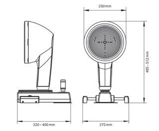
| Boyutlar (G x D x Y) | 275 x 320 - 400 x 485 - 512 mm |
| Ağırlık | 6,1 kg |
| Maks. güç tüketimi | 18 W |
| Voltajı | 90 - 264 VAC |
| Frekans | 47 - 63 Hz |
| Önerilen bilgisayar özellikleri | İşlemci: Intel® Core i5-7600 veya üstü, 8 GB RAM, Sabit sürücü alanı: 1 TB, Windows® 10 Pro |
| Önerilen ekran çözünürlüğü | 1920 x 1200 piksel |
In acc. with the Medical Device Directive 93/42/EEC

The OCULUS QM system is certified in accordance with ISO 13485 and (EU) 2017/745 (MDR).

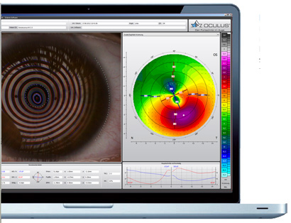
JENVIS Pro ile kuru göz sendromunun nedenini hızlı ve güvenilir bir şekilde öğrenin. Keratograph 5M'de bulunan yeni JENVIS Pro Dry Eye Report, mevcut teşhis ve tedavi sürecinizde size yardımcı olacaktır. Kuru göz sendromunu teşhis etmek için ölçüm sonuçlarını temel alarak kapsamlı bir tarama yapın. Tüm sonuçlar, Tıbbi Ürünler Yasasına uygun şekilde belgelenir ve hastanız için düzenli ve kolay anlaşılır bir çıktı halinde özetlenir.
Keratograph 5M'de sunulan yeni JENVIS Pro Dry Eye Report etkili tarama, sağlam temelli ölçüm sonuçları ve hasta memnuniyeti gibi tüm avantajlarından yararlanın.
Kuru göz sendromu için tarama yapmak, her refraksiyonun rutin bir parçası olmalıdır.
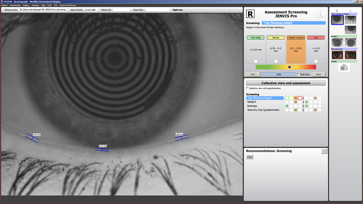
Dört parçadan oluşan tarama testi, gözyaşı filmi miktarı ve kalitesindeki anormallikleri hızlı ve doğru bir şekilde ortaya çıkarır.5 dakika süren bu tarama işlemi, her refraksiyondan önce rutin olarak gerçekleştirilmelidir. JENVIS Pro gözyaşı filmi tarama rutini; gözyaşı menisküs yüksekliği ölçümü, gözyaşı filmi kırılma (break-up) zamanı, bulbarda kırmızılık ve kısa bir anket içerir.
Hastanızda kuru göz sendromu olup olmadığına hızla ve kolayca karar verin.
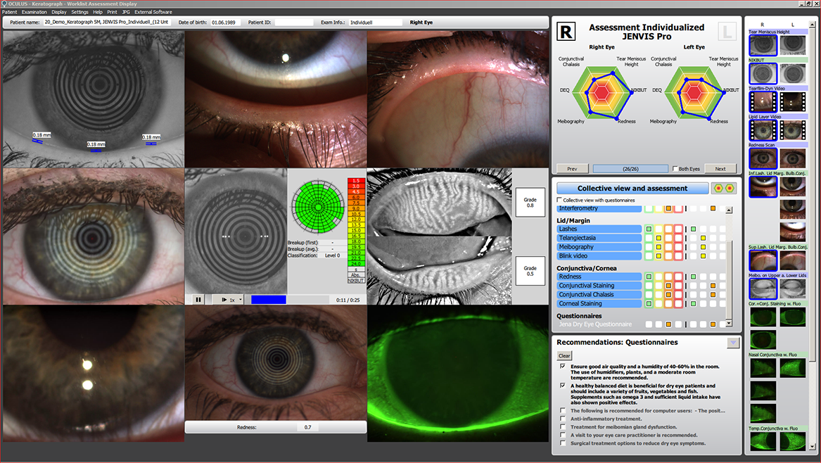
JENVIS Pro Dry Eye Report ile kapsamlı kuru göz sendromu analizi için gerekli tüm değerlendirme kriterlerini karşılayan, hatalara karşı korumalı bir test dizisine sahip olursunuz. Yararlı ipuçları ve ölçümlerinizde size destek sağlayacak optimum, önceden tanımlanmış ayarlar sunarak kuru göz sendromu için hızlı ve etkili analizler gerçekleştirebilmenizi ve ön segment bulgularınızı belgeleyebilmenizi sağlar.
Hastalarınızın kuru göz semptomlarını hafifletmek için onlara özel tavsiyeler verebileceğiniz donanımlar edinin.
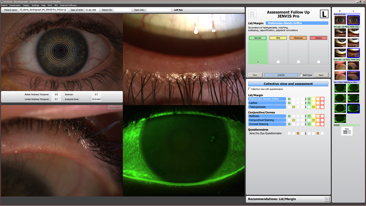
Tedavi önerilerinizin başarılı olup olmadığını bilmek mi istiyorsunuz? Problemin temel nedenini tanımlamak için ayar seçeneklerini kullanarak takip işlevinden yararlanın. Böylece hastanızın gözyaşı filmindeki iyileşmeleri başarıyla belgeleyebileceksiniz.
Düzenli kontroller, hastalarınızın gözünde kurtarıcı bir uzman olmanızı sağlayacaktır.
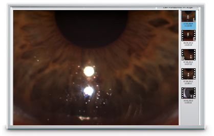
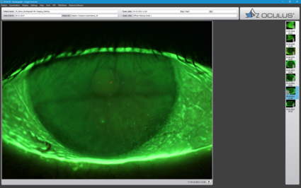
Gözyaşı filmi beyaz veya kızıl ötesi aydınlatma kullanılarak değerlendirilir. Yeni yüksek çözünürlüklü renkli kamera, en küçük yapıları bile görünür kılar. NIKBUT'a (Non-invazif Keratograph Kırılma Süresi) ve gözyaşı menisküs ölçümüne ek olarak, TF-Tarama lipid katmanının ve gözyaşı filmi dinamiklerinin değerlendirmesini de yapabilir. OCULUS Keratograph 5M ile gözyaşı filmi analizi non-invaziftir ve herhangi bir ek araç olmadan yürütülür.
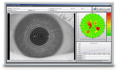
Gözyaşı filmi kırılma süresi non-invazif olarak ve tamamen otomatik bir şekilde ölçülür.. Yeni kızıl ötesi aydınlatma insan gözü tarafından görünür değildir. Bu, muayene sırasında gler olmasını önler. TF-tarama, sonuçları sizin ve müşterilerinizin kolayca anlayacağı bir şekilde sunar.
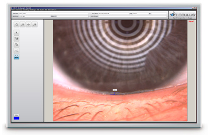
Yırtık menisküsün yüksekliği entegre bir cetvel ve çeşitli büyütme seçenekleriyle hassas bir şekilde ölçülebilir ve alt gözkapağı kenarı boyunca gelişimi değerlendirilebilir. Sonuçlar, hasta dosyasına kaydedilir.
Lipid katmanının ve yapısının interferans renkleri görünür hale getirilebilir ve kaydedilebilir. Lipid katmanının kalınlığı yapıya ve renge göre değerlendirilir.
Saniyede 32 çerçeveye kadar çıkabilen video kaydı, gözyaşı filmi partikül akışının gözlemlenmesini sağlar ve bu sayede gözyaşı filminin viskozitesiyle ilgili sonuçlar çıkarılabilir.
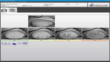
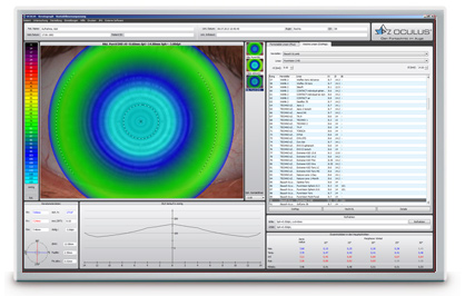
Keratograph 5M'nin çok işlevselliği, meibografi gibi zor muayenelere kolayca ve etkili bir şekilde entegre olur. Meibomian bezlerinin disfonksiyonu kuru gözün en yaygın sebebidir. Bez dokusundaki morfolojik değişiklikler, Meibo-Tarama kullanarak görünür hale getirilebilir ve JENVIS Meibo Derecelendirme Ölçekleri ile sınıflandırılabilir.
Görüntüleme Yazılımı, video ve görüntü dosyalarını kaydetmek için kullanılır. Videoları ve tekli görüntüleri izlemenin yanında, ayrıca kayıtları muayene edilen gözün simüle edilmiş floresein görüntüleriyle karşılaştırabilirsiniz.
Sonuç: Statik floresein görüntüler ve videolar biyomikroskop koşulları altında kaydedilebilir!
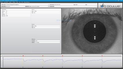
OxiMap®, lens dioptrisine göre yumuşak kontakt lenslerin oksijen geçirgenliğinin renk haritasını temsil eder, bu da kolayca anlaşılabilir - müşterileriniz tarafından bile!
Oksijen geçirgenliği (Dk/t), kontakt lensin yapıldığı materyale ve onun kalınlığına bağlıdır. Geçmişte, sadece -3.0 dpt kontakt lens merkezindeki oksijen geçirgenliği hakkında üreticilerin spesifikasyonları bulunuyordu. OxiMap® sayesinde şimdi lens kalınlığına göre tüm alan üzerindeki Dk/t değerlerinin grafik görselleştirilmesini elde edebiliyorsunuz.
Dk/t değerleri renk kodlamasına sahiptir, siyah renk kapalı bir gözde bulunandan daha düşük oksijen beslemesini temsil eder. Kontakt lensleri takarken korneanın bütünlüğünü korumak için, kontakt lenslerin takılacağı süreye göre minimum Dk/t değerleri önerilir. OxiMap® renk kodlaması bu uluslararası önerilere dayanmaktadır.
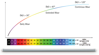
OxiMap® lens dioptrisine göre ayrı olarak ayarlanır ve hastanız için en uygun kontakt lensi seçmenize yardımcı olur. Yumuşak kontakt lensler için kullanılan yeni materyaller mükemmel oksijen geçirgenliği sağlar.
OxiMap® JENVIS Araştırma ve Jena Uygulamalı Bilimler Üniversitesi ile işbirliği içinde geliştirilmiştir.

OCULUS Dünyası
Sıcak Gündem
Ürün Kategorileri
|
OCULUS kalite yönetim sistemi, ISO 13485 ve (AB) 2017/745 (MDR) standartlarına göre sertifikalandırılmıştır. |